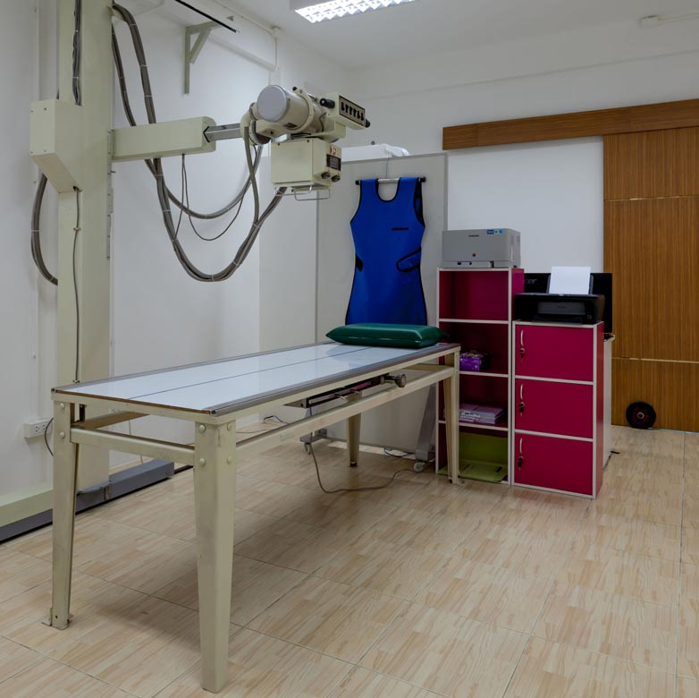
Digital Imaging X-ray
X-rays use radiation to create an image of inside the body, at First Western, we use Digital Imaging X-rays which are more advanced and faster to produce results than the traditional X-ray. Bones are very hard and dense which means that they show up very clearly on an X-ray, therefore, X-rays are most commonly used to diagnose bone-related issues. Although blood vessels and organs are not so clear on an X-ray, they are visible; therefore a chest X-ray can help examine the heart, major arteries and lungs. Some of the problems that X-rays help identify include:
• Breaks or fractures
• Dental problems
• Thinning or weakening of the bones (osteoporosis)
• Bone infections (osteomyelitis)
• Abnormal spinal curvatures (scoliosis)
• Benign or malignant bone tumours
• Heart conditions
• Lung conditions
Types of X-ray include:• Dental problems
• Thinning or weakening of the bones (osteoporosis)
• Bone infections (osteomyelitis)
• Abnormal spinal curvatures (scoliosis)
• Benign or malignant bone tumours
• Heart conditions
• Lung conditions
Barium enema – this procedure involves barium solution being pumped into the bowel and can be used to diagnose such as chronic constipation or blood in the faeces.
Angiography- is a type of X-ray used to create images of blood vessels called angiograms which show blood vessels. A dye is first injected into the area which helps X-ray ‘see’ the soft tissues.
To help make the blood vessels show up, a contrast medium is injected into the appropriate area. A contrast medium (dye) highlights the blood vessels as it travels through them, problems like blockages can be identified as the way in which the dye moves through the blood vessels is studied.
Intravenous urogram (IVU)- can be used to diagnose problems with the urinary system. During an IVU, a contrast medium such as iodine solution will be administered by injection into your veins and move into your kidneys and bladder thus enabling X-rays of your urinary system to be taken.
What to expect during an X-ray
A typical X-ray procedure usually takes around 20 minutes. Depending on which part of your body needs the X-ray, you may be asked to put on a hospital gown in private and remove any jewellery.
Once inside the X-ray room, your radiographer will either help you get into the correct position or you may be asked to sit on a chair or lie down on an X-ray table so that an image of the appropriate part of your body can be captured. Your radiographer will ask you to stay still and may ask you to hold a deep breath for a moment in the case of a chest exam.
The scanner will be operated from a control room. He or she will be able to see, hear and speak to you at all times during the procedure. Once the scan is complete, the scanner table will move back out and you will be helped down. You will usually be able to go home as soon as you feel ready. The radiographer will be operating from behind a screen but you will be able to hear each other speak at all times. They may ask you to move into different positions in order to gather more images from different angles. If this is difficult for you then you will be assisted.
The results will soon be reviewed by our radiologists who specialise in using medical images to make diagnoses and you will usually hear your results within X days.